Mole or skin-tag removal at a med spa: identify the lesion before destroying it
A cosmetic label such as “mole” or “skin tag” is not a diagnosis. Before laser, plasma, cautery, freezing, shaving, or snipping, establish who identified the lesion, whether tissue should be preserved for pathology, and who owns the result and follow-up.
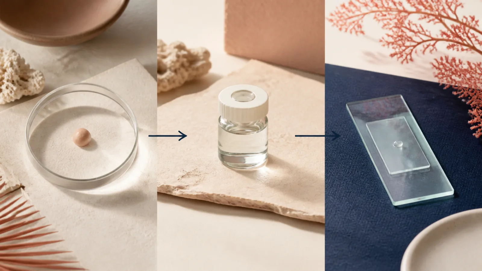
A spot should be identified before it is cosmetically destroyed. “Mole,” “skin tag,” and “benign growth” can describe appearance, not a confirmed diagnosis. The useful preprocedure record names the person who examined it, the working diagnosis, why removal is appropriate, whether a specimen will exist, whether pathology is indicated, and who will deliver and act on the result.12
This sequence matters because vaporizing, burning, freezing, or chemically destroying tissue can leave no intact specimen. A later photograph cannot always answer the question a pathologist could have addressed from preserved tissue.
The first decision is identity, not equipment
| What the marketing calls it | What still needs to be established | Why the answer changes the plan |
|---|---|---|
| Skin tag | Whether the growth is consistent with a skin tag or is a look-alike such as a wart or skin cancer | A look-alike may need a different method, no cosmetic treatment, or diagnostic review |
| Mole | Whether it is a stable benign nevus or has features that warrant biopsy | Destroying a suspicious lesion can eliminate tissue and delay a diagnosis |
| Uncertain growth | Whether a qualified clinician can identify it or diagnostic sampling is needed | The answer determines whether cosmetic destruction, specimen-preserving removal, or referral is appropriate |
Standardized photographs are useful documentation, but they do not convert a cosmetic assessment into pathology. Ask whether the evaluation included size, color, border, surface, symptoms, change over time, prior treatment, personal history, and the surrounding skin.
Removal and biopsy are different jobs
A dermatologist may remove a lesion by shave technique or surgical excision and send tissue for microscopic examination.1 A skin tag may be snipped, frozen, or treated with electrosurgery after the clinician has identified it.2 The same physical action can serve a diagnostic, therapeutic, or purely cosmetic purpose depending on the lesion and specimen plan.
Laser, intense light, plasma arc, radiofrequency, cautery, cryotherapy, acids, and topical products destroy tissue in different ways. Their presence on a menu does not establish that a device is appropriate for a particular lesion. The plasma-device guide explains why a shared marketing word does not establish the device, indication, or operator.
Ask the proceduralist to name the method literally:
- excision with closure;
- shave removal with a specimen;
- snip removal;
- cryotherapy;
- electrosurgery or cautery;
- laser or another energy device; or
- a topical or chemical product.
Then ask what tissue remains after that method and what would cause the plan to change before treatment begins.
A pathology plan has four owners
“We can send it out” is incomplete. A reliable chain identifies the person who orders the examination, the laboratory that receives the specimen, the clinician who interprets the report in context, and the person responsible for communicating next steps.
If no pathology is planned, the record should say why the clinician considers destruction reasonable and what change would trigger referral or biopsy instead. “It looks fine” is not a durable handoff.
A title does not establish diagnostic authority
The person advertising the service may use “aesthetician,” “laser specialist,” “skin expert,” or another private credential. Those titles do not show a medical license, scope, supervision, or authority to diagnose a lesion. Verify the individual in the applicable state record, the business or facility record when relevant, and the professional who owns the diagnostic decision.
In Florida, start with the license-verification guide. Match the legal name and license number to the person who will examine and treat the spot. Do not substitute a medical director’s name for the operator’s identity.
The product deserves the same precision. FDA warning letters have addressed sellers marketing unapproved mole- and skin-tag-removal drug products.3 An ingredient name, dietary-supplement label, “natural” claim, or marketplace listing is not evidence that a finished product is approved for removing skin lesions.
Cosmetic success and diagnostic completeness are separate
A lesion can look flatter while pigment or cells remain. It can recur after a partial removal. A scar can be smaller than the original spot yet still matter aesthetically. Conversely, complete excision may create a longer line than a superficial shave. No single method minimizes recurrence, preserves pathology, and guarantees the smallest mark for every lesion.
Ask the clinician to separate:
- the diagnostic goal;
- the tissue-removal goal;
- the cosmetic scar goal;
- the recurrence plan; and
- the follow-up trigger.
This prevents a promise such as “scar-free” from obscuring the decision about specimen depth or margins.
Do not let price erase the result pathway
Normalize the estimate by lesion count, body site, evaluation, anesthesia, procedure, pathology, dressings, suture removal, result visit, and any staged work. A low per-lesion price may omit the diagnostic visit or pathology charge; a higher quote does not prove that either is included.
Preserve the consent, photographs, product or device name, settings when applicable, specimen record, pathology report, wound-care instructions, and follow-up communications. If a concern later needs routing, the reporting guide separates urgent evaluation, professional complaints, product reports, and consumer issues.
Use a lesion-first booking sequence
- Name the spot without assuming the diagnosis. Record the location, duration, change, symptoms, prior treatment, and standardized photographs.
- Identify the diagnostic owner. Verify who examines the lesion, their active license and role, and when a dermatology or other medical evaluation occurs.
- Choose the method after identity. Ask what tissue the proposed shave, excision, freezing, cautery, laser, plasma, or topical product removes or destroys.
- Decide the specimen path in advance. Document whether tissue will be preserved, where it goes, what test is ordered, and why pathology is or is not planned.
- Assign result ownership. Name who reviews the report, how it is delivered, and what happens if the diagnosis, margins, or sample are uncertain.
- Reconcile appearance and follow-up. Set expectations for scar, pigment change, recurrence, wound care, and the findings that require reassessment.
The decisive question is: “Who established what this lesion is, and will this removal method preserve the diagnostic evidence and follow-up path that the answer may require?”
Sources
- American Academy of Dermatology. Moles: Diagnosis and treatment. Dermatologist examination, biopsy when a mole could be cancerous, removal methods, and pathology review. Accessed .
- American Academy of Dermatology. Skin tags: Why they develop, and how to remove them. Look-alike lesions, in-office removal methods, and the absence of FDA-approved at-home skin-tag removal products. Accessed .
- U.S. Food and Drug Administration. Warning Letter: Ariella Naturals. FDA action addressing products marketed with disease and mole- or skin-tag-removal claims as unapproved drugs. Accessed .