DEXA vs InBody vs 3D body scan: tracking body composition
DEXA estimates bone mineral and soft tissue using low-dose X-rays; InBody-style bioimpedance estimates composition from electrical impedance; a 3D optical scan measures surface shape and predicts circumferences or composition. Their numbers should not be mixed into one trend line.
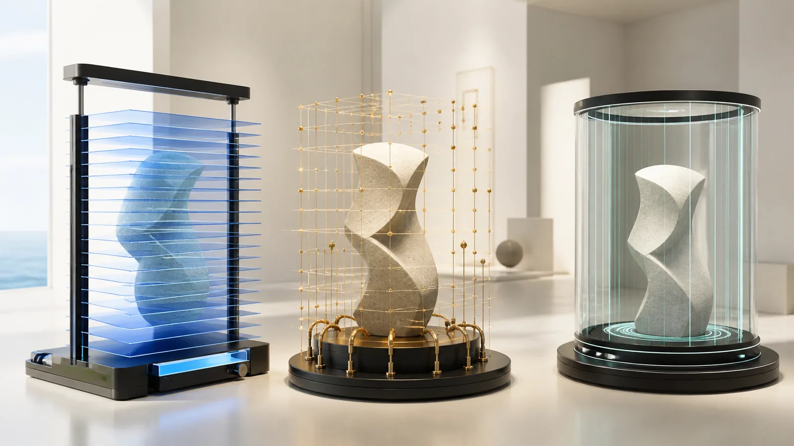
DEXA, bioelectrical impedance, and 3D optical scanning estimate different physical properties. DEXA uses low-dose X-rays to model bone mineral and regional lean and fat tissue; InBody and other BIA devices infer body composition from electrical impedance and body-water models; 3D scanners capture surface shape and use algorithms to estimate circumferences, volume, or composition. The outputs are not interchangeable. 1234
Choose the method that matches the question, then repeat on the same device under comparable conditions. Switching systems can create a false “change” larger than the biological change being tracked.
The machines do not directly see “body fat” the same way
Every method combines measurement with assumptions. DEXA measures X-ray attenuation and partitions pixels into bone mineral and soft-tissue estimates. BIA measures impedance and applies equations involving body water and demographic inputs. A 3D scanner measures the external surface, then predicts internal composition using a model trained against reference data.
| Dimension | DEXA / DXA | BIA / InBody-style | 3D optical scan |
|---|---|---|---|
| Physical signal | Low-dose X-ray attenuation | Electrical impedance through body segments or whole body | Visible/infrared surface geometry |
| Strong use | Bone density; regional and total soft-tissue estimate on some systems | Fast repeat measurements under standardized conditions | Circumference, volume, shape and visual change tracking |
| Key sensitivity | Machine, software, positioning, region definitions, body size and hydration | Hydration, food, exercise, bladder, temperature, skin contact and equation | Clothing, pose, breathing, landmarks, camera calibration and prediction model |
| Exposure | Small ionizing-radiation exposure | No ionizing radiation | No ionizing radiation |
| Main caution | Not a direct four-compartment chemical analysis | Consumer decimal precision can exceed biological accuracy | Surface precision does not guarantee composition validity |
| Trend rule | Repeat same system and positioning | Repeat same model, protocol and hydration context | Repeat same scanner, pose, clothing and software version |
“Gold standard” is often used too loosely. DXA is an important reference method and clinical tool, especially for bone density, but body-composition estimates still depend on hardware, software and assumptions. A four-compartment model can answer composition differently by combining multiple measurements, as one 3D validation study demonstrated. 4
DEXA: regional detail with radiation and positioning
RadiologyInfo, produced by ACR and RSNA, describes DXA as a quick, low-dose X-ray exam used primarily to measure bone loss and fracture risk. 1 Whole-body systems can also report lean and fat estimates by region, but a wellness scan should not be confused with a diagnostic bone-density interpretation.
Ask whether the appointment is a clinical bone-density study, a whole-body composition scan, or both; which regions and outputs are reported; who interprets it; and whether the scanner and software are approved and maintained for that use. Pregnancy or possible pregnancy needs to be disclosed because the method uses ionizing radiation.
Positioning, motion, metal, clothing, recent contrast studies, body size, and software-defined region boundaries can affect results. A “visceral fat” output may be an estimate rather than a direct image of every internal fat depot. Request the device, software version, precision information, and least significant change used by the facility.
Frequent scanning should have a reason. Low dose is not zero dose, and tiny short-term changes may be smaller than measurement error or normal hydration and glycogen shifts.
BIA: accessible, repeatable, and hydration-sensitive
BIA sends a small electrical current and measures resistance and reactance; equations estimate total body water and derive fat-free and fat mass. Multi-frequency and segmental systems can add detail, but brand, electrodes, posture, equations and populations differ.
An independent study of 15 BIA devices found substantial variability in validity and in the ability to track change, illustrating why two devices cannot be treated as interchangeable scales. 2 A high repeatability score does not prove that the value equals a reference method; it may mean the same system produces a stable estimate under similar conditions.
Standardize time of day, food and drink, recent exercise, alcohol, bladder, skin temperature, menstrual context, and contact. Follow the device’s instructions rather than inventing a dehydration protocol. Dehydrating to produce a “better” percentage compromises the meaning of the result.
InBody is a brand, not the name of the entire method. Record exact model and software. A gym’s older foot-to-foot system and a clinic’s segmental multi-frequency device may produce different values even on the same day.
3D scanning: strong shape data, modeled composition
Optical systems can create a body mesh and automate circumferences and volumes. This makes them useful for visualizing shape change, clothing fit, waist or hip trends, and regional volume without radiation.
In a multiethnic validation study of a commercial scanner, measurements showed high test-retest precision and strong agreement with criterion measures for several outputs, but offsets and limits of agreement remained. 3 Another study found acceptable agreement with DXA for some composition estimates but poor validity against a four-compartment model and an average overestimate of body-fat percentage. 4 Both can be true: a model can track consistently and still differ from another method.
Ask which exact scanner and algorithm version is used, what population trained the prediction equation, whether the output is directly measured or inferred, and whether software updates reprocess old scans. Shape data should not be marketed as a medical diagnosis or proof of visceral-fat change without appropriate validation.
Privacy is also distinctive: a body mesh or near-nude image can be sensitive. Ask about clothing protocol, local versus cloud processing, raw-image retention, model training, access, exports, deletion, and whether the visual is used in marketing.
Match method to the job
For clinical bone density, a properly ordered and interpreted DXA has a distinct role. For regional composition under a consistent clinical protocol, whole-body DXA may be useful. For frequent low-burden trend feedback, BIA can be practical when conditions are standardized. For shape, circumference and visual geometry, 3D optical scanning can be the clearest match.
For a small change over a few weeks, waist tape, strength, clothing, symptoms, adherence, and other outcomes may be as useful as a body-fat decimal. The test should not become the goal.
Avoid clinics that use a proprietary “metabolic age,” body score, or visceral rating without defining formula, reference population, error, and decision relevance. A colorful report does not make an estimate diagnostic.
Software can move the baseline without moving the body
These systems turn a physical signal into an estimate through software. A firmware update, new prediction equation, changed demographic field, revised segmentation boundary, or replacement machine can shift results even when the person has not changed. Ask the operator to record versions and whether older scans are recalculated after an update.
Export the full report, not only a dashboard screenshot. Keep raw or primary outputs that the system permits, date and time, device serial or location, posture and protocol, and the facility’s explanation of each field. If an upgrade breaks continuity, close the old series and establish a new baseline rather than applying a homemade correction factor.
Cross-site comparison is particularly weak when franchises use different models under one brand name. Confirm hardware at each address before buying a package that promises portable progress tracking.
Design a trend that survives noise
- 1. Define the decision Choose bone health, regional composition, hydration-sensitive trend, circumference, shape or another outcome; do not scan simply to collect every number.
- 2. Record the system Save manufacturer, model, software and algorithm version, operator, positioning, clothing and report definitions.
- 3. Standardize conditions Repeat at a similar time with comparable food, fluid, exercise, bladder, temperature, pose and recent-treatment context.
- 4. Respect method limits Ask for precision error, least significant change, validation population, direct versus inferred outputs and pregnancy or radiation considerations.
- 5. Keep one trend line Do not subtract BIA from DXA or 3D from BIA. Start a new baseline after a device or major software change.
- 6. Pair numbers with useful outcomes Track waist, strength, function, clothing, symptoms, adherence and clinical markers appropriate to the goal instead of optimizing a single percentage.
The most trustworthy body-composition trend is deliberately boring: same question, same machine, same conditions, enough time, and no attempt to make incompatible estimates agree.
Sources
- RadiologyInfo.org. Bone densitometry (DEXA, DXA). ACR/RSNA patient reference used for DXA's low-dose X-ray mechanism, bone-density role, preparation, radiation, limitations, and pregnancy questions. Accessed .
- PubMed Central. Assessing the reliability and cross-sectional and longitudinal validity of fifteen bioelectrical impedance analysis devices. Independent comparison used for device-to-device BIA variability, cross-sectional agreement, longitudinal change, and the need to keep device and conditions consistent. Accessed .
- PubMed. Assessment of clinical measures from a commercial 3-dimensional optical body scanner. Multiethnic validation study used for 3D optical scan precision, agreement with DXA and manual measures, offsets, and limits of agreement. Accessed .
- PubMed. Validity of a 3-dimensional body scanner against a 4-compartment model and DXA. Independent validation used to show that acceptable agreement with DXA did not equal validity against a four-compartment criterion and that method choice changes the answer. Accessed .