Mammograms after breast augmentation, lift, reduction, or fat transfer: prepare the record
Prior breast surgery does not create one replacement screening schedule. Tell the imaging facility about implants, lift, reduction, fat transfer, scars, symptoms, and prior imaging before the appointment; keep routine cancer screening distinct from imaging ordered to evaluate an implant or a new finding.
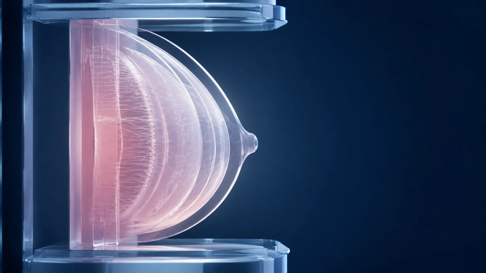
After breast augmentation, lift, reduction, or fat transfer, continue breast-cancer screening on the schedule set for the person’s age and risk unless the responsible clinician changes it; prior surgery does not create one universal substitute schedule.5 Tell the imaging facility in advance about every breast operation, implant and graft record, symptoms, and prior images, and keep routine screening separate from imaging ordered to evaluate implant integrity or a new clinical finding.1367
The practical job is continuity: getting the right history and comparison studies to the interpreting team without trying to choose the imaging test from an article.
Three imaging questions should not be collapsed
| Question | Typical trigger | Record that clarifies it |
|---|---|---|
| Breast-cancer screening | Age- and risk-based preventive plan without a new symptom | Screening recommendation, risk history, prior images and reports |
| Diagnostic breast evaluation | New lump, pain, skin or nipple change, discharge, asymmetry, or an imaging finding needing workup | Symptom location and timing, clinical exam, prior surgery and imaging |
| Implant-integrity evaluation | Product-specific surveillance plan or concern for rupture or another implant complication | Implant type, device card, operation date, symptoms, FDA and clinician guidance |
A mammogram can contribute to more than one question, but the indication determines the protocol and follow-up. The ACR implant guideline organizes imaging choices by implant type, symptoms, age, and suspected complication rather than offering one “implant scan.”3
Call the facility before scheduling
Tell the scheduler whether the breasts contain saline or silicone implants, whether surgery involved a lift, reduction, reconstruction, explant, capsule operation, or fat transfer, and whether there is a new symptom. Ask whether the site regularly images people with that history and whether it needs extra appointment time.
For implants, FDA advises informing the mammography facility so technologists can use implant-displacement views when appropriate.1 The history helps the team plan positioning and explain limitations. It does not mean the person should skip mammography or assume every facility uses the same protocol.
Verify that the site is certified under the Mammography Quality Standards Act through FDA’s facility resources. MQSA gives patients rights concerning a written summary and access or transfer of mammography records.2 Accreditation, certification, an order, and an interpreting radiologist’s qualification are separate records; ask the facility when any of them is unclear.
Bring an operation map, not just a procedure label
“Breast surgery” leaves out the details an imaging team may need. Build a concise history:
- every breast procedure and date;
- surgeon and facility;
- side and incision pattern;
- implant manufacturer, model, surface, fill, size, lot or serial when available, and pocket location;
- lift or reduction technique and pathology report;
- fat-transfer donor and recipient areas, date, and staged sessions;
- explant, capsule, revision, infection, biopsy, radiation, or cancer history;
- current symptoms and exact location; and
- prior mammography, ultrasound, MRI, biopsy, and operative images or reports.
If a device card is missing, request the operative report and implant log from the surgeon or facility. Do not guess the fill or model from feel or a scar.4
Implants can change technique and have their own follow-up
FDA notes that breast implants can obscure breast tissue on mammography, which is why additional implant-displacement views may be used.1 FDA has also received reports of suspected implant rupture during mammography and says the extent of the problem is unknown because reporting is limited; that uncertainty belongs in informed planning rather than a promise of zero risk.1 Tell the technologist about pain, firmness, suspected rupture, or a recent operation before positioning begins.
Silicone-implant surveillance is a different job from cancer screening. FDA product labeling and clinician guidance may call for ultrasound or MRI to look for silent rupture, while a mammogram addresses breast tissue and can reveal other findings. The MRI-versus-ultrasound guide explains that product-specific pathway. One test does not silently replace the other.
Lift and reduction create a different history
A lift repositions the breast envelope; a reduction also removes tissue and ordinarily generates a pathology record. Either can leave scars and internal changes that are useful for the radiologist to know.6 Obtain the operative and pathology reports and preserve preoperative and postoperative imaging.
If the surgeon recommends a preoperative mammogram, ask whether the purpose is routine screening, risk-based assessment, a symptom, or surgical planning—and who will review it before surgery. “Medical clearance” should never mean that an unread report follows the patient into the operating room.
After surgery, do not choose an interval based on a generic online healing rule. Ask the breast-imaging and surgical teams when the planned examination can be performed comfortably and safely, and which clinician owns any delay.
Fat transfer can generate findings that need context
Fat grafting moves a person’s own fat into breast tissue. Subsequent palpable or imaging findings may require comparison with the operation record and prior studies.7 The existence of fat transfer should not be used to self-label a new lump as harmless fat necrosis.
Give the imaging team dates, treated regions, volumes if documented, staged sessions, and any postoperative infection, drainage, injury, or prior biopsy. A new or changing finding belongs with the responsible clinical team; it should not wait for a routine screening appointment simply because surgery occurred.
Close every loop in writing
After imaging, obtain the patient summary and full report. Confirm who received it, whether prior studies were available for comparison, whether additional imaging or clinical evaluation is recommended, and who orders and tracks that next step. If records move to a new facility, follow MQSA procedures and confirm arrival rather than relying on a portal request alone.2
- Separate the indication. State whether the appointment is routine screening, diagnostic workup, implant surveillance, or more than one job.
- Notify the facility early. Describe implants and every lift, reduction, fat-transfer, reconstruction, biopsy, or revision before the visit.
- Transfer the comparison record. Send prior images and reports, operation and pathology reports, and device information; confirm receipt.
- Name any symptom precisely. Record side, location, onset, change, skin or nipple findings, pain, swelling, trauma, and relevant surgery date.
- Assign result ownership. Know who reads the report, communicates it, orders follow-up, and confirms completion.
The decisive scheduling question is: “What imaging job are we doing, what surgery and device records does this facility need, and which clinician owns the result and every recommended next step?”
Sources
- U.S. Food and Drug Administration. Breast Implant Adverse Events During Mammography. FDA information used for implant disclosure, mammography technique, reported rupture events, and the distinction between screening and implant concerns. Accessed .
- U.S. Food and Drug Administration. Frequently Asked Questions About MQSA. Current FDA mammography-quality guidance used for facility certification, reports, record transfer, and patient access. Accessed .
- American College of Radiology. ACR Appropriateness Criteria: Breast Implant Evaluation. Imaging guideline used to keep asymptomatic implant surveillance, suspected complication, implant type, age, and cancer evaluation as separate imaging questions. Accessed .
- U.S. Food and Drug Administration. What to Know About Breast Implants. FDA overview used for device records, ongoing follow-up, screening, and symptom-reporting context. Accessed .
- U.S. Preventive Services Task Force. Breast Cancer: Screening. Current national screening recommendation used to anchor routine screening as an age- and risk-informed clinical plan rather than an implant-surveillance schedule. Accessed .
- Breast Cancer. What we should know in mammography after reduction mammoplasty and mastopexy?. Clinical imaging study used for characteristic mammographic changes after reduction mammoplasty and mastopexy and the need to preserve the surgical context. Accessed .
- Plastic and Reconstructive Surgery. Mammographic changes after fat transfer to the breast compared with changes after breast reduction: a blinded study. Blinded comparative study used for the existence and interpretation of post-procedure mammographic findings after breast fat transfer and reduction. Accessed .