Ablative vs nonablative vs fractional laser: the terms are not opposites
Ablative and nonablative describe what laser energy does to tissue; fractional describes how treatment is distributed in microscopic columns. A laser can therefore be fractional ablative or fractional nonablative, and the exact device and settings still matter.
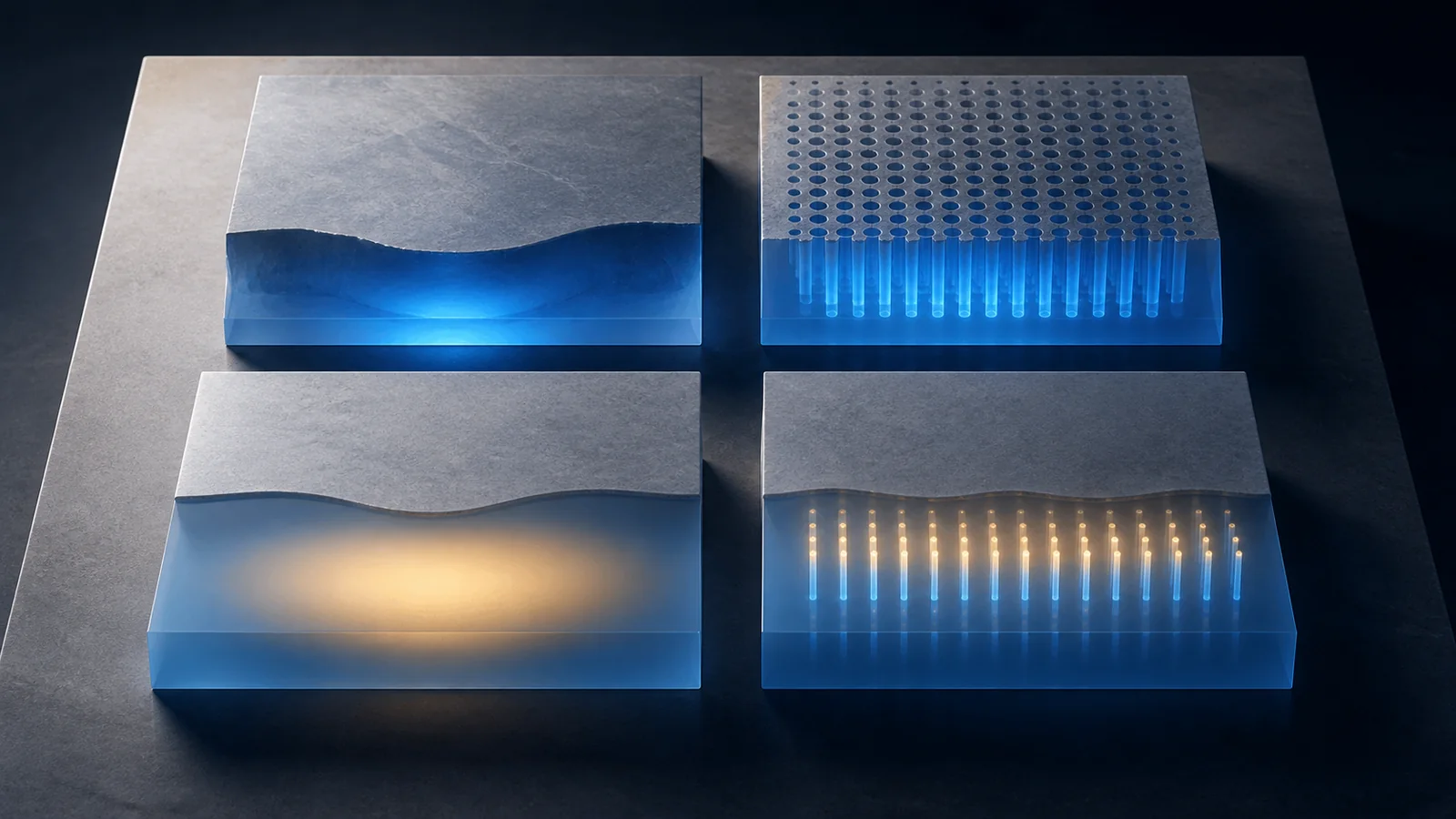
Ablative and nonablative are tissue-effect categories; fractional and full-field are delivery patterns. Ablative energy removes microscopic or continuous areas of tissue. Nonablative energy heats or coagulates targeted tissue while leaving the surface largely intact. Fractional treatment spaces microscopic treatment zones among untreated tissue, so it can be either ablative or nonablative.
That two-axis model resolves a common menu error: “fractional” is not automatically a gentle alternative to “ablative.” A fractional CO2 treatment can be ablative, while a fractional erbium-glass treatment can be nonablative.12
Put every laser into a two-axis matrix
| Tissue effect | Full-field delivery | Fractional delivery |
|---|---|---|
| Ablative | Removes or vaporizes a continuous treated surface or field | Creates separated microscopic columns of ablation with intervening tissue |
| Nonablative | Heats a broader target while preserving the surface barrier | Creates separated microscopic zones of thermal injury or coagulation beneath a largely intact surface |
The matrix is only the first layer. Wavelength, pulse duration, fluence or energy, spot size, density, passes, cooling, treatment depth, overlap, anatomy, and skin context can make two sessions in the same box meaningfully different.
Ablative describes removal, not a brand
Common ablative resurfacing systems use wavelengths strongly absorbed by water, including CO2 and erbium:YAG categories. The energy vaporizes or removes targeted tissue and produces surrounding thermal effects that depend on wavelength and settings.3 Full-field ablation treats a continuous surface; fractional ablation creates columns.
Ask how much surface remains untreated, the intended depth, density, number of passes, and whether the plan involves only fractional delivery or includes confluent treatment in selected areas. A product name such as “CO2” does not reveal those parameters.
Because the barrier is disrupted where tissue is ablated, wound care, infection prevention, pigment response, sun avoidance, and recovery supervision become central. The provider should define when re-epithelialization is expected, which changes are normal, and which require prompt evaluation without promising an exact recovery for every person.
Nonablative describes surface preservation, not no recovery
Nonablative resurfacing directs thermal injury below or through a preserved surface to stimulate a repair response. Fractional nonablative systems create microscopic thermal zones rather than removing columns.2 The intact surface can reduce open-wound recovery compared with an ablative exposure, but redness, swelling, pigment change, blistering, infection, or scarring can still occur depending on the device and plan.
“No downtime” is not a technical category. Ask what the practice means: no open wound, ability to work next day, or no visible redness. Those are different endpoints.
Fractional describes coverage, not intensity
Fractionation leaves intervening tissue between treatment zones. Density describes how much of the area is covered; depth and energy describe what happens within each zone. Raising one parameter is not interchangeable with raising another.
FDA clearance K244060, for example, identifies fractional and nonfractional handpieces and exact ablation and resurfacing indications for one CO2 system.1 K221770 describes a nonablative fractional erbium-fiber system.2 The records prove why a clinic must name the model; they are not comparative endorsements.
Match the target before debating strength
Texture, fine lines, pigment, vascular change, scars, and laxity are not one laser target. Diagnose the target in the consultation rather than selecting from device branding. Existing pigment tendencies, melasma, active inflammation, infection risk, prior isotretinoin or procedures, medication, sun exposure, history of abnormal scarring, and ability to perform aftercare can change the plan.
Comparative evidence does not support a simple “ablative always wins” or “nonablative is always safer” rule. One meta-analysis found small and heterogeneous randomized samples and no clear categorical superiority across its pooled facial and hand rejuvenation outcomes.4 Results depend on indication, device, protocol, outcome measure, and follow-up.
Compare treatment burdens on the same horizon
One deeper session and a series of lower-disruption sessions create different calendars. Compare:
- the exact endpoint and how it will be measured;
- total planned sessions and reassessment points;
- numbing, medications, supplies, and follow-up included;
- days of wound care versus days of visible redness or swelling;
- sun and activity restrictions in South Florida conditions;
- probability of needing maintenance or another modality; and
- who evaluates delayed pigment change or an unexpected response.
Do not equate fewer scheduled sessions with lower total burden. Recovery support and inability to tolerate sun exposure can be the controlling constraint.
Make the review protocol as specific as the treatment
Laser proposals often devote more detail to the procedure than to the evidence that will be used afterward. That reverses the order of a sound comparison. Before treatment, record the exact concern, its location, and the conditions under which it is visible. Texture, pigment, a scar edge, and a fine line need different photographs and may need different follow-up intervals. A broad promise to make skin look “better” cannot be audited.
Create three separate dates rather than one vague results appointment:
- an early recovery check for barrier healing, swelling, discomfort, and the aftercare plan;
- a defined outcome review after transient redness or surface change is less likely to dominate the image; and
- a later decision point for maintenance, another session, or a different modality.
At each review, repeat camera, lens distance, lighting direction, expression, makeup status, and head position. Record whether the image is meant to show healing, pigment, texture, or contour. Do not compare a dry, unretouched baseline with a moisturized or differently lit follow-up. If a clinic uses software scoring, ask which feature the score measures and whether the same capture settings are locked at both visits.
This discipline also protects against category-level conclusions. The comparative literature combines different devices, protocols, outcomes, and follow-up periods, which is one reason a simple ablative-versus-nonablative winner is difficult to establish.4 A result from one fractional density, depth, and skin context does not validate every treatment carrying the same category name.
A useful chart therefore connects target → device and settings logic → recovery checkpoint → outcome measure → next decision. If any link is missing, the proposed “strength” is not yet a complete treatment plan.
Verify one proposal in order
- Name the clinical target. Ask what feature is being treated and how change will be measured at a stated follow-up.
- Place the plan in the matrix. Identify ablative or nonablative effect and fractional or full-field delivery.
- Open the device record. Match manufacturer, model, handpiece, wavelength, and intended use to the proposed area and goal.
- Translate the settings logic. Ask how depth, energy, density, passes, cooling, and anatomy are chosen without trying to prescribe settings yourself.
- Map recovery and response. Get written aftercare, expected barrier and visible recovery, direct clinical contact, and a path for urgent or delayed concerns.
The resurfacing-versus-peel-versus-microneedling guide helps choose a treatment family. This article solves the next decision inside laser resurfacing. A precise proposal should fit in one sentence: named device, named handpiece, ablative or nonablative effect, fractional or full-field delivery, defined target, and recovery plan.
Sources
- U.S. Food and Drug Administration. 510(k) indications for use K244060. Official example distinguishing fractional and nonfractional handpieces and ablation, resurfacing, and coagulation indications for an exact CO2 platform. Accessed .
- U.S. Food and Drug Administration. 510(k) summary K221770. Official example of a nonablative fractional erbium-fiber laser intended for fractional skin resurfacing and soft-tissue coagulation. Accessed .
- PubMed. Ablative laser resurfacing. Peer-reviewed clinical review used for tissue-target and fractional-column definitions, common laser categories, evaluation, and complications. Accessed .
- PubMed. Systematic review and meta-analysis comparing ablative and nonablative lasers. Systematic review supporting caution about categorical superiority claims and highlighting heterogeneity and small comparative samples. Accessed .
- U.S. Food and Drug Administration. Medical lasers. FDA consumer and provider overview of medical-laser intended uses, labeling, operation, and hazards. Accessed .