Breast thermography vs mammogram: why heat imaging is not a screening substitute
Breast thermography records surface heat; mammography uses low-dose x-rays to image breast tissue. FDA says thermography has not been shown effective as a standalone breast-cancer screen and cleared uses are adjunctive. A normal thermogram cannot replace a recommended mammogram or explain a symptom.
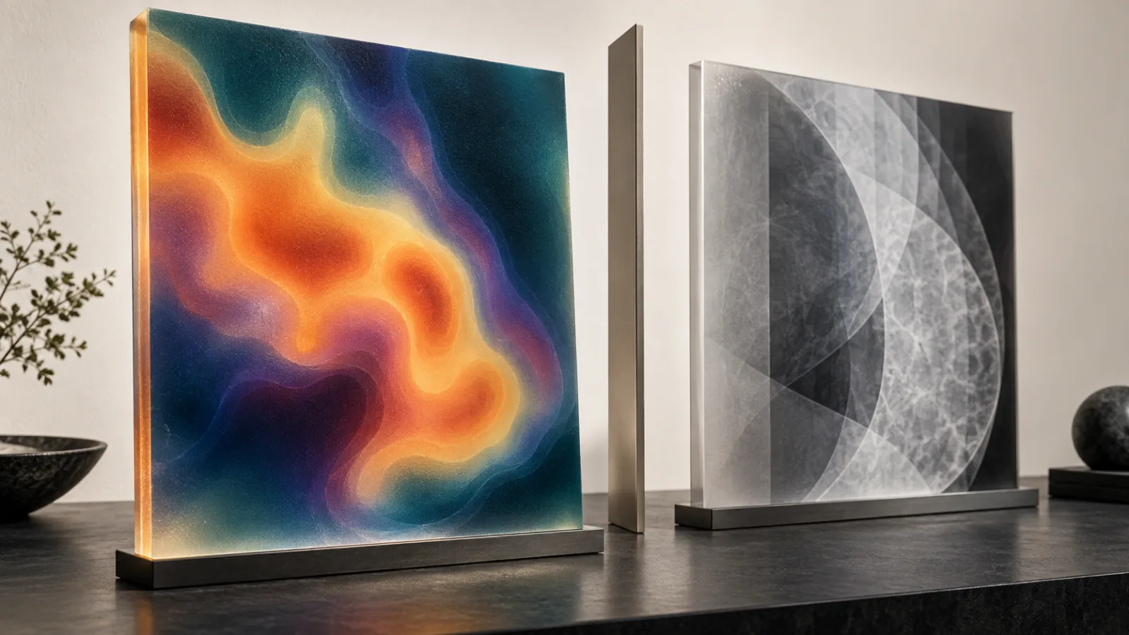
Breast thermography and mammography measure different things. Thermography maps infrared surface-temperature patterns; mammography uses low-dose x-rays to image internal breast tissue. FDA says thermography has not been shown effective as a standalone breast-cancer screening test and that cleared thermography devices are adjunctive. A normal thermogram cannot replace a recommended mammogram or evaluate a new breast symptom by itself.12
This answer does not set an individual’s screening schedule. Age, sex assigned at birth, symptoms, pregnancy, prior cancer or high-risk lesions, genetics, family history, breast density, implants, prior radiation, and other factors can change the appropriate pathway. The important consumer distinction is that avoiding compression or x-rays does not make a different signal an equivalent screen.
Heat patterns are not tissue images
| Dimension | Thermography | Mammography |
|---|---|---|
| Signal | Infrared radiation from skin surface translated into temperature patterns | Low-dose x-ray attenuation through breast tissue |
| Primary regulatory role | Adjunctive use for specifically cleared devices and labeling | Established breast-cancer screening and diagnostic imaging roles |
| What it cannot do alone | Reliably rule out cancer or replace standard screening | Guarantee no cancer; some findings need additional imaging or biopsy |
| Physical experience | No breast compression for image capture | Breast positioning and brief compression |
| Main marketing trap | “No radiation” becomes “better or earlier detection” without comparative outcome evidence | A normal result is treated as a permanent all-clear |
Temperature can vary with inflammation, hormones, environment, vascular patterns, technique, acclimation, skin conditions, and other factors. An abnormal heat pattern is not a cancer diagnosis; a symmetric or “cool” pattern is not proof that internal tissue is normal.
FDA clearance must be read as adjunctive
FDA warns that no valid scientific data show thermography is an effective standalone screening tool for detecting breast cancer early, and notes that devices may be cleared only for use with another test such as mammography.1 “FDA cleared” should therefore be followed by the exact device name, 510(k) number, intended use, and required accompanying method.
Ask the thermography provider:
- exact device and FDA record;
- whether the label says adjunctive;
- who interprets the images and their credentials;
- what preparation and room controls are used;
- what a positive, negative, or indeterminate classification means;
- how the report reaches the clinician who owns breast care;
- whether the provider explicitly tells clients not to delay mammography or diagnostic evaluation.
A device registration, facility listing, operator certificate, or “medical-grade camera” does not expand the intended use.
Mammography has benefits and limitations
Mammography is the only modality for which population screening recommendations and mortality evidence are established in the current USPSTF framework.3 It uses ionizing radiation and can yield false positives, false negatives, additional imaging, biopsy, and overdiagnosis; breast density can reduce sensitivity.4 Those limitations are reasons for informed screening and quality systems, not proof that thermography is equivalent.
FDA administers the Mammography Quality Standards Act program for mammography facilities.2 Verify a facility through the current FDA/MQSA record. If a consumer service offers “thermal breast screening” outside that system, clarify that it is not mammography and does not inherit MQSA status.
Screening, diagnosis, and implant surveillance are separate jobs
Screening is intended for people without a known breast symptom in a defined population. A lump, discharge, focal skin change, persistent pain, swelling, or new contour change requires clinical evaluation; buying a thermogram is not a diagnostic shortcut.
Implant-integrity imaging answers another question. FDA recommendations for ultrasound or MRI surveillance of certain silicone implants do not replace breast-cancer screening. The implant MRI-versus-ultrasound guide separates shell integrity from cancer detection.
After augmentation, lift, reduction, or fat transfer, keep operative reports and device information and tell the imaging facility before the appointment. The mammography-after-breast-surgery guide covers positioning, prior images, scar and fat-grafting records, and implant-displacement views.
“Earlier” requires outcome evidence
Thermography marketing may claim that heat or vascular change appears years before a tumor becomes visible. To evaluate such a claim, request prospective evidence in the intended screening population comparing the complete thermography pathway with the current standard. The study should predefine thresholds, blind interpretation, confirm outcomes, report interval cancers, and show whether earlier detection improves clinically meaningful outcomes rather than merely moving the diagnosis date forward.
Testimonials and before-and-after thermal palettes cannot establish sensitivity, specificity, stage shift, false-positive burden, or mortality benefit. Color-map intensity is a visualization choice, not proof of disease severity.
Image-acquisition controls do not solve the screening-evidence gap
Thermography protocols may specify room temperature, acclimation, distance, camera calibration, positioning, recent exercise, topical products, caffeine, smoking, sun exposure or timing relative to procedures. These controls can improve repeatability of a surface-temperature image. They do not transform the method into a validated standalone cancer screen.
Ask whether interpretation is automated, performed by a physician, or outsourced; what training or credentials are claimed; and whether the interpreter has access to history and prior breast imaging. A proprietary score should disclose its validation population, threshold, false-positive and false-negative performance, and how software changes are controlled.
If serial thermograms are compared, preserve the original files and protocol metadata. A changed palette, scale, crop or threshold can make the same temperature field look different. “Stable” thermal images also cannot override a new symptom or an abnormal mammogram.
An abnormal thermogram still needs a conventional pathway
Before buying the test, ask what happens after a red, asymmetric, high-risk or indeterminate report. Does the service arrange clinical examination and appropriate diagnostic imaging, transfer the original data, and track resolution—or simply recommend that the customer contact another clinician?
The downstream clinician should know that the trigger was thermography and should choose the next step based on symptoms, risk and established breast-imaging pathways, not a thermal color alone. Repeating the thermogram until it looks reassuring can delay evaluation; proceeding straight from a heat map to biopsy without conventional assessment can create a different harm.
Compare convenience without substituting purpose
Thermography may be comfortable, quick, and radiation-free. Those are service attributes. They do not answer whether the method detects the clinically important target accurately enough to replace mammography. A mammogram’s compression or radiation can be discussed with a qualified imaging clinician; fear-based claims that imply the standard itself causes more cancer than it detects require strong evidence.
Price comparisons should include interpretation, report transfer, repeat imaging, additional mammographic or ultrasound workup, and medical consultation. A thermography subscription that still requires standard imaging is an add-on cost, not a replacement saving.
The decisive question
Ask: “Is this exact thermography device cleared only as an adjunct, and what evidence shows it can replace—not merely accompany—the screening or diagnostic pathway recommended for me?” Under current FDA guidance, it should not be marketed as a standalone substitute for mammography.
Sources
- U.S. Food and Drug Administration. Breast cancer screening: thermogram no substitute for mammogram. FDA's current distinction between adjunctive thermography clearance and standalone mammographic screening. Accessed .
- U.S. Food and Drug Administration. Mammography. FDA overview of mammography, MQSA, benefits, limitations, radiation, and facility requirements. Accessed .
- U.S. Preventive Services Task Force. Breast cancer: screening. Current population-level mammography recommendation and evidence boundaries. Accessed .
- National Cancer Institute. Breast cancer screening (PDQ)—health professional version. Evidence review for breast screening modalities, benefits, false results, overdiagnosis, and harms. Accessed .