Gynecomastia: liposuction vs gland excision starts with fat, gland, skin, and cause
Liposuction removes accessible fat; excision directly removes firm gland or other tissue and can address skin or the nipple-areola complex. Many operations combine them. Before technique, confirm what tissue is present and whether a medical cause or suspicious finding needs its own evaluation.
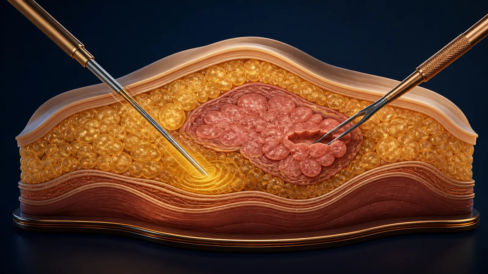
Gynecomastia liposuction and gland excision are not competing brand packages. Liposuction removes fat that a cannula can aspirate; excision directly removes firm glandular tissue and can address excess skin or nipple-areola anatomy. Many male-breast-reduction operations combine both. The technique decision should follow an examination that distinguishes fat, gland, skin, asymmetry, and any medical or suspicious finding.12
A flat-price “gland removal” package can be just as incomplete as “scarless lipo.” The first may not define how much tissue remains under the nipple or how the surrounding chest is blended; the second may not address firm gland or loose skin.
Put each tissue in its own row
| Finding or goal | Technique it may call for | Question the label cannot answer |
|---|---|---|
| Diffuse fatty fullness | Liposuction may reduce accessible subcutaneous fat | Whether true gland, skin excess, or another mass is also present |
| Firm tissue under the areola | Direct excision may be needed for tissue not removable by cannula | How much should remain to support a smooth nipple-areola contour |
| Fat plus firm gland | A combined operation can contour the chest and directly remove resistant tissue | Which step does what and where each incision will be |
| Loose or redundant skin | Skin excision or a staged plan may be discussed | Whether skin will contract enough after volume removal alone |
| Puffy, enlarged, low, or asymmetric areola | A procedure-specific areola or skin plan may be added | Whether the proposed scar and blood-supply design fit the anatomy |
| Discrete, hard, eccentric, fixed, or otherwise concerning finding | Clinical and possibly imaging or tissue-diagnosis pathway before cosmetic design | That the finding is ordinary gynecomastia |
ASPS describes liposuction, excision, and combination techniques rather than one universal operation.1 Ask the surgeon to mark fat, palpable gland, skin excess, chest borders, and asymmetry on a diagram. “Grade” systems may summarize severity, but the marked anatomy is more useful for comparing two plans.
Cause evaluation is not cosmetic gatekeeping
Gynecomastia can be associated with normal hormonal stages, medicines or substances, endocrine or systemic conditions, or no identified cause. The EAA guideline recommends a history and physical examination directed at the breast and genitalia, with additional testing selected for the clinical context.2
Bring a complete medication and supplement record, including hormones, anabolic agents, hair-loss products, psychiatric or cardiac medicines, cannabis and other substances, and dates relative to change. Do not stop a prescribed drug based on a cosmetic consultation. The responsible clinician can decide whether a suspected contributor has an appropriate alternative.
Duration and stability matter. So do pain, nipple discharge, rapid or one-sided change, a distinct mass, skin or nipple retraction, lymph-node findings, and personal or family history. These facts do not diagnose the cause; they determine whether the surgical sales path should pause for a different evaluation.
Imaging is selected by the finding, not ordered automatically
The ACR imaging framework treats a physical examination typical of gynecomastia differently from an indeterminate mass or an examination suspicious for malignancy.3 That means neither “everyone needs a mammogram” nor “men never need breast imaging” is a sound clinic rule.
If imaging or a prior biopsy exists, obtain the actual report and, when relevant, images and pathology. A screenshot of “normal” is not a full baseline for a surgeon who must plan around a persistent or changing finding.
“Gland excision” still requires a contour plan
Removing every palpable bit beneath the areola may create a depression, tethering, adherence, or edge. Leaving too much can leave fullness. The consultation should define the intended residual layer, how the transition to the surrounding chest will be blended, and what the surgeon will do if the left and right sides differ.
Likewise, liposuction needs specifics:
- areas treated and feathered;
- cannula access points and likely scars;
- whether a power, ultrasound, laser, or other assisted system is proposed;
- wetting solution and anesthesia plan;
- expected aspirate versus the visible tissue goal; and
- the threshold for adding direct excision.
Device or technique branding does not prove superior contour. The broader liposuction comparison explains why surgical fat removal is a different exposure from a noninvasive device.
Skin response should be discussed before “minimal scar” promises
Younger, elastic skin may redrape differently from stretched or redundant skin, but no photograph can guarantee contraction. Ask what skin the surgeon expects to remain, where folds might persist, whether a larger excision is an immediate or staged option, and what scars each route creates.
Prior weight change, chest surgery, scars, asymmetry, sun damage, stretch marks, nicotine exposure, and a history of raised scars can alter planning. If revision is staged, define the decision date, expected evidence of settling, and who pays for the second operation.
Tissue handling and pathology need an answer
Directly excised tissue creates a specimen. Ask whether it will be sent for pathology, how it will be labeled by side and site, who receives the result, and how an unexpected result is communicated. A clinic should not improvise this after surgery.
Pathology cannot make liposuction aspirate equivalent to an oriented excision specimen. Nor does a routine pathology result retroactively prove that no preoperative assessment was needed. The cosmetic lesion pathology guide explains the general principle: destruction or removal technique affects what can later be evaluated.
Compare complete operations
ASPS lists risks including contour irregularity, asymmetry, changes in sensation, fluid accumulation, poor wound healing, tissue injury, scarring, and possible revision.4 Translate each material risk into the surgeon’s prevention, recognition, and response plan rather than comparing complication lists in the abstract.
A complete chest-contouring map labels each finding—fat, gland, skin, or another mass—and assigns it a step, incision, specimen decision, and contingency. That map makes a technique comparison meaningful and exposes any part of the anatomy the proposed plan does not address.
Sources
- American Society of Plastic Surgeons. Gynecomastia Surgery Procedure Steps. Professional description of liposuction, excision, and combined techniques in male breast reduction. Accessed .
- European Academy of Andrology. EAA Clinical Practice Guidelines—Gynecomastia Evaluation and Management. Guideline addressing history, examination, underlying causes, imaging or laboratory evaluation when indicated, and management principles. Accessed .
- American College of Radiology. ACR Appropriateness Criteria: Evaluation of the Symptomatic Male Breast. Evidence-based imaging framework distinguishing typical gynecomastia from an indeterminate or suspicious male breast finding. Accessed .
- American Society of Plastic Surgeons. Gynecomastia Surgery Risks and Safety. Procedure-specific consent topics for contour, healing, sensation, asymmetry, fluid, tissue loss, and revision. Accessed .